Sunday, 7 June 2020

Thursday, 4 June 2020
Monday, 3 February 2020
Diagnosis of CUP - carcinoma of unknown primary
Definition of Carcinoma of Unknown Primary (CUP):

- No obvious identifiable primary site, despite a careful clinical history, physical examination, radiologic imaging, and biochemical or histologic investigations.
- Immunohistochemistry plays a vital role in diagnosis and classification of CUP lesions.
- Most common cases of CUPs --> carcinoma.
- Adenocarcinoma accounts for ~70% cases of CUPS.
- Poorly differentiated carcinoma: 15-20%
- Squamous cell carcinoma 5%
- Neuroendocrine carcinoma 5%
Steps:
1. To find the line of differentiation - lineage markers --> keratins (Pancytokeratin/ CAM 5.2), lymphoid (LCA), melanoma (S100), germ cell (OCT 3/4), and sarcoma markers (Vimentin).
2. To determine the type of CK distribution in the tumor cells - some subsets of CKs are seen in certain tumours.
3. To see if there is coexpression of vimentin.
4. To see if there is expression of supplemental antigens of epithelial or germ cell derivation, that
is, carcinoembryonic antigen (CEA), epithelial membrane antigen (EMA), or placental alkaline
phosphatase (PLAP).
5. Finally look for expression of cell-specific products, cell-specific structures, and transcription
factors or receptors that are unique identifiers of cell types.

Ref: Dabb's Diagnostic immunohistochemistry
Endomyocardial Biopsy
EMB - Endomyocardial Biopsy
- Widely used for surveillance of cardiac allograft rejection and unexplained ventricular dysfunction.
- Should be done in young patients with myocarditis and older patients with infiltrative cardiac disease.
Approach:
Through femiral vein or artery
Right internal jugular vein (preferred)
Biopsy samples are taken from IVS (interventrucular septum) - since RV wall is thin and can lead to perforation.
Can be guided by fluoroscopy or 2D echocardiography.
Transthoracic echo guidance is preferred.
Commonly used bioptomes:
1. Novatome
2. Argon EMB forceps
3. Bipal 7 bioptome.
Five biopsies are taken and put in isotonic saline.
Transferred to
1. 10% neutral buffered formalin - rejection, cardiomyopathy, myocarditis abd tumours
2. Zeus fixative - for IF studies for rejection (C4d)
3. 4% glutaraldehyde - electron microscopy for drug toxicity, metaboloc or storage disease and LCDD (light chain deposition disease)
4. Snap frozen tissue in liquid nitrogen - PCR (viral myocarditis) Dystrophin (muscular dystrophy)
EMB is absolutely necessary for diagnosis of the following conditions:
1. Anthracycline induced cardiomyopathy - loss of myofilaments and vacuolar degeneration.
2. Cardiac allograft rejection
3. Sarcoidosis
4. Giant cell myocarditis
5. Hylereosinophilic syndrome
Complications of EMB:
IMMEDIATE:
Site hematoma
Transient RBBB
Transient arrhythmias
Tricuspid regurgitation
RV perforation -- cardiac tamponade and pericardial effusion
Occult pulmonary embolism
LATE:
Coronary artery to RV fistula formation
Severe tricuspid regurgitation
TENSINS - A short note
TENSINS:
Integrins are a family of proteins which form important connections between the cell and the ECM(extracellular matrix).
Focal adhesions are generated following interaction of integrins with ECM.

Functions of focal adhesions:
Tensin gene: Has 4 members
1) TNS1 - Normally localised to Heart, skeletal muscle, kidney and lung
2) TENC1
3) TNS3 - Normally localised to Placenta, kidney
4) TNS4/ CTEN - Normally localised to Placenta, prostate
------------------------------------------------------------------------------------------------------------
Structure of Tensin:
Has 2 domains: Cytoskeletal domain (N-terminus) and signal transduction domain (C-terminus).
I) Cytoskeletal domain (N terminus)
Composed of two Actin Binding Domains (ABD) and PTEN
The cytoskeletal domain is absent in cten.
II) Signal transduction domain (C terminus)
Composed of Src-homology2 (SH2 domain) and PTB (Phosphotyrosin binding domain)


Actin is also required for cell migration. But after a certain point, more than the strength of the actin pull, it is the strength of adhesion that is inhibitory to movement.
Cancer cell dissemination occurs by 2 strategies - 1. Amoeboid movement as single cells 2. Movement in cell clusters.
Amoeboid movement is facilitated by cten.

- Tensin is a cytoplasmic phosphoprotein.
- Localized to integrin-mediated focal adhesions.
Integrins are a family of proteins which form important connections between the cell and the ECM(extracellular matrix).
Focal adhesions are generated following interaction of integrins with ECM.
- They come to the site of action through integrins.
- Include numerous proteins (approximately 60) and include:
- Talin
- Tensin
- Vincullin
- Paxillin
- Src
- Focal adhesion kinase (FAK)
- Protein Kinase C

Functions of focal adhesions:
- Important in the cytoskeletal organisation and signal transduction.
- These focal adhesions in general are important in cell adhesion, migration, differentiation, proliferation, tissue development, apoptosis and genesis of disease.
Coming back to Tensins:
- Tensins act as a molecular bridge between integrins and actin cytoskeleton.
- It is a 220KD cytoplasmic phosphoprotein.
- Localised at focal adhesins.
- Phosphorylates on tyrosin, serine and threonine residues.
Tensin gene: Has 4 members
1) TNS1 - Normally localised to Heart, skeletal muscle, kidney and lung
2) TENC1
3) TNS3 - Normally localised to Placenta, kidney
4) TNS4/ CTEN - Normally localised to Placenta, prostate
------------------------------------------------------------------------------------------------------------
Structure of Tensin:
Has 2 domains: Cytoskeletal domain (N-terminus) and signal transduction domain (C-terminus).
I) Cytoskeletal domain (N terminus)
Composed of two Actin Binding Domains (ABD) and PTEN
The cytoskeletal domain is absent in cten.
- ABD interacts with actin filaments
Composed of Src-homology2 (SH2 domain) and PTB (Phosphotyrosin binding domain)
- SH2 interacts with tyrosine phosphorylated and non-phosphorylated proteins and leads to signal transduction.
- PTB interacts with the cytoplasmic tails of β integrin.

------------------------------------------------------------------------------------------------------------
Role of tensins in health and disease:
Play an important role in
1. Cell adhesion and motility
2. Cell survival and apoptosis
1. Cell adhesion and motility
Cten and tensin 3 control the migratory phenotype by direct interaction with integrin.
Resting state -- tensin 3 stabilises the actin-beta integrin complex.
On EGF stimulation -- cten competes with tensin 3 to bind to integrin resulting in loss of interaction with integrin, weakening of adhesion and cell migration.
Resting state -- tensin 3 stabilises the actin-beta integrin complex.
On EGF stimulation -- cten competes with tensin 3 to bind to integrin resulting in loss of interaction with integrin, weakening of adhesion and cell migration.

Cell migration --> promotion of metastasis in cancers.
Invasion of cancer cells is thus facilitated by EGF dependant switch in isoforms of tensin.
Invasion of cancer cells is thus facilitated by EGF dependant switch in isoforms of tensin.
It is also important in the pathogenesis of wound healing, gastrulation etc where migration of cells occur.
Mutations of KRAS/BRAF/MAPK (as seen in many tumours) -> upregulation of cten --> metastasis.
Actin is also required for cell migration. But after a certain point, more than the strength of the actin pull, it is the strength of adhesion that is inhibitory to movement.
Cancer cell dissemination occurs by 2 strategies - 1. Amoeboid movement as single cells 2. Movement in cell clusters.
Amoeboid movement is facilitated by cten.
2. Cell survival and apoptosis:

Detachment of integrins --> Loss of contact with the extracellular matrix --> Cell death
Tensin and its downstream signaling molecules may be targets for therapeutic interventions in wound healing and cancer.
Tuesday, 26 March 2019
Prostatic carcinoma - Rules!!
Rule 1: Prostatic carcinoma is suspected always on low power examination.
Rule of "Toos" (at low magnification):
1. Too small glands
2. Too crowded glands (with back to back arrangement)
3. Too clear glands

Three criteria for confirmation of malignancy (at high magnification):
1. Enlarged nuclei
2. Prominence of nucleoli
3. Loss of basal cells
Additional features that indicate a malignancy:
1. Crystalloids
2. Glomerulations
3. Circumferential perineural invasion
Figure below - Shows benign prostatic acini with basal cells and luminal cells


Additional features that indicate a malignancy:
1. Crystalloids
2. Glomerulations
3. Circumferential perineural invasion
Figure below - Shows benign prostatic acini with basal cells and luminal cells

Figures below - Prostatic adenocarcinoma


Friday, 22 March 2019
Hamartoma versus Choristoma
Hamartoma:
Disorganised but benign mass composed of cells indigenous to the involved site.
Choristoma:
Heterotopic rest of normal cells.
Disorganised but benign mass composed of cells indigenous to the involved site.
Choristoma:
Heterotopic rest of normal cells.
Biphasic Soft tissue tumours with epithelial component
1. Chondroid syringoma / mixed tumour of soft tissue
2. Malignant Peripheral Nerve Sheath Tumour
3. Synovial Sarcoma
2. Malignant Peripheral Nerve Sheath Tumour
3. Synovial Sarcoma
Thursday, 21 March 2019
Tuesday, 19 March 2019

Tuesday, 5 March 2019
Jones silver methanamine stain
Jones silver methanamine stain
Used to highlight basement membranes.
Used as a routine stain in renal biopsies.
Do you remember the principle of PAS stain? This one is similar to that.
JONES STAIN:
C-C bonds in carbohydrates (Eg:Glycogen) ----> Oxidised by periodic acid ----> Aldehyde ---> Reduces silver solution ----> Metalllic silver (Black)
PAS STAIN:
C-C bonds in carbohydrates (Eg:Glycogen) ----> Oxidised by periodic acid ----> Aldehyde ------> Schiff's reagent (Fuschin-sulphurous acid) ------> Alkyl sulphonate (magenta coloured compound).

Fig: Black lines (arrows) represent the glomerular and tubular basement membranes.
Arrow heads - Capillary walls.
Note: It is not specific for basement membranes, demonstrates any carbohydrate which can be oxidised to aldehyde.
PAS STAIN:
C-C bonds in carbohydrates (Eg:Glycogen) ----> Oxidised by periodic acid ----> Aldehyde ------> Schiff's reagent (Fuschin-sulphurous acid) ------> Alkyl sulphonate (magenta coloured compound).
- Structures that are stained magenta on a PAS stain will be black in Jones' stain.

Fig: Black lines (arrows) represent the glomerular and tubular basement membranes.
Arrow heads - Capillary walls.
Note: It is not specific for basement membranes, demonstrates any carbohydrate which can be oxidised to aldehyde.
Friday, 8 February 2019
What is haematoxylin?
HAEMATOXYLIN:
- Derived from the wood Haematoxylum campechianum.
- It means bloodwood - refers to the dark red colour (Haemato - blood, xylon- wood)
- Campechianum refers to the coastal city of Campeche on the Yucatan Peninsula, the locality of the heart wood.

Pic: Google

- Haematoxylin has little or no staining capacity.
- Haematoxylin is oxidised to haematein which gives the blue colour.
- Oxidation - two methods - 1. Natural 2. Chemical
- If we start with hematein, oxidation will start to diminish the staining capacity right from the start, shortening the working life of the solution ☺☺
Mucormycosis - How to identify?
Mucormycosis
- Transplant recipients
- Uncontrolled diabetes mellitus
- Patients with hematological malignancies
- Immunosuppressed patients
- People living with HIV (PLHIV)
HOW TO IDENTIFY MUCORMYCOSIS:
Broad, aseptate fungal hyphae, branching at 90 degree.
Angioinvasion is frequently seen, leading to thrombosis of vessels and tissue infarction & necrosis.
Mucor is always seen as a basophilic "glassy" structure on H&E staining.

PAS and Silver Methanamine stain highlight the fungal hyphae.

Pic: PAS stain

Silver methanamine: Arrow points to the blood vessel, fungal hyphae are seen infiltrating the wall.

DIFFERENTIAL DIAGNOSIS:
ASPERGILLUS:
Slender, septate hyphae with acute angle branching.
Note: Candida species is often seen with Mucor and Aspergillus.
Monday, 28 January 2019
Helicobacter pylori - More than half of world's population is affected, including you and me probably!!!
Helicobacter pylori
Helicobacter pylori causes the following:
1. Gastritis
2. Gastric ulcers
3. MALToma
4. Gastric carcinoma.
How do we identify H.pylori gastritis?
Many of us would have been taught to look for H.pylori organisms in a gastric biopsy with neutrophils (foveolitis/ foveolar abscess/ lamina propria infiltrates).
This is true!!!..
However, H.pylori is the most common type of CHRONIC GASTRITIS.
Presence of neutrophils only signifies active inflammation and likely presence of organisms in the biopsy.
Signs of H.pylori gastritis:
- Loss of mucin in the surface epithelium and epithelial damage leading to irregularity of the surface.
- Chronic inflammation in the lamina propria with lymphoid aggregates and plasma cells.
How do they look?
H.pylori are 2-4um curved structures.
Where to find them?
- Surface mucin
- Adherent to surface foveolar cells
- Within the foveolar pits

In figure, green circles - indicate plasma cells.
Curved organisms seen within the red circles - Helicobacter pylori.
Differential diagnosis for H.pylori:
Helicobacter heilmannii - twice as long as H.pylori and has 5-7 spirals.
Wednesday, 16 January 2019
Mönckeberg medial calcific sclerosis - An interesting finding!!
- First described by Johann Georg Monckeberg.
- It occurs most often in patients >50 years of age.
- Calcium deposits are seen in the muscular wall (media) of blood vessels.
- Results in thickening of the arterial wall and loss of elasticity.
- Does not have any serious pathologic consequence, since the lumen is patent.

In figure, arrow points to purple fragmented deposits that represent calcium.
The special stain that would highlight calcium is Von Kossa (stains the deposits black).
Monday, 14 January 2019
"Are You Afraid Of Monster Cells?" - Seminal Vesicle
Seminal vesicle
- Tall columnar epithelial cell lining with a layer of basal cells.
- Presence of golden brown refractile granules in the cytoplasm - LIPOFUSCIN.
- Lipofuscin - Also found in the ampulla of vas deferens and prostatic portion of ejaculatory ducts.
Important and an unusual feature: Presence of MONSTER CELLS - Cells with enlarged, hyperchromatic, irregularly shaped nuclei.
- Do not interpret these cells as malignant cells!!!
- May also be seen in the ampulla of vas deferens.


In figure, arrow points to the pigment; circle points to the MONSTER CELLS.

Arrow - MONSTER CELLS.
It is important to recognise seminal vesicle epithelium for two reasons:
1. Infiltration of seminal vesicle epithelium by prostatic carcinoma (pT3b).
2. To avoid misinterpretation of the seminal vesicle cells (monster cells) as malignant cells in core biopsies of prostate.
Sunday, 13 January 2019
Why is identifying ganglion cells important?
Ganglion cells are absent in Hirschsprung disease (also called aganglionic megacolon).
Anatomy:
There are two neural plexus in the intestine - Submucosal (Meissner's) and myenteric (Auerbach's).
Submucosal (Meissner's) plexus - Present just beneath the muscularis mucosa.

In figure, red arrow --> Muscularis mucosa, blue arrow --> muscularis propria.
Space between the two (green line) --> submucosa; Yellow circle --> Submucosal plexus of ganglion cells.
Myenteric (Auerbach's) plexus - Present inbetween the two muscular layers of the bowel wall.

In figure, arrow --> neuronal processes
Circle --> Ganglion cell clusters
Important points:

In figure, arrow --> neuronal processes, circle --> ganglion cells.

Diagnosis of Hirschsprung disease:
Absence of ganglion cells in the submucosal and the myenteric plexus.
Other features in favour of Hirschsprung's disease:
Hypertrophy of nerve bundles.
Hypertrophy of muscularis mucosae.
Anatomy:
There are two neural plexus in the intestine - Submucosal (Meissner's) and myenteric (Auerbach's).
Submucosal (Meissner's) plexus - Present just beneath the muscularis mucosa.

Space between the two (green line) --> submucosa; Yellow circle --> Submucosal plexus of ganglion cells.
Myenteric (Auerbach's) plexus - Present inbetween the two muscular layers of the bowel wall.

Circle --> Ganglion cell clusters
Important points:
- Ganglion cells are often associated with neuronal processes and Schwann cells.
- These interconnect and create a neural network throughout the submucosal layer.
- Often occur in clusters and may mimic giant cells, epithelioid cells or granulomas.

In figure, arrow --> neuronal processes, circle --> ganglion cells.

Diagnosis of Hirschsprung disease:
Absence of ganglion cells in the submucosal and the myenteric plexus.
Other features in favour of Hirschsprung's disease:
Hypertrophy of nerve bundles.
Hypertrophy of muscularis mucosae.
Thursday, 27 December 2018
Ganglion cells - They will find you, before you find them😀
Ganglion cells are neuronal cell bodies.
They are identified by their ....


They are identified by their ....
- Large size
- Polygonal shape
- Large eccentrically placed vesicular nuclei
- Prominent eosinophilic nucleolus
- Abundant eosinophilic cytoplasm


Saturday, 22 December 2018
Pilocytic astrocytoma - Squash preparation
Pilocytic Astrocytoma:

Most striking feature: Hair like (pilocytic processes) seen in the background.
Cells are monomorphic (uniform in size and shape) with ovoid bipolar bland appearing nuclei.
Many Rosenthal fibres and Eosinophilic Granular Bodies will be seen.
Rosenthal Fibres:

Eosinophilic Granular Bodies:

For those of you who are still thinking about "What a squash preparation is?"
- Most common astrocytoma in children.
- Most common sites: Cerebellum, Floor of 3rd ventricle, thalamus, hypothalamus.

Most striking feature: Hair like (pilocytic processes) seen in the background.
Cells are monomorphic (uniform in size and shape) with ovoid bipolar bland appearing nuclei.
Many Rosenthal fibres and Eosinophilic Granular Bodies will be seen.
Rosenthal Fibres:
- Brightly eosinophilic, thick, glassy cork-screw shaped/ carrot shaped srtuctures.
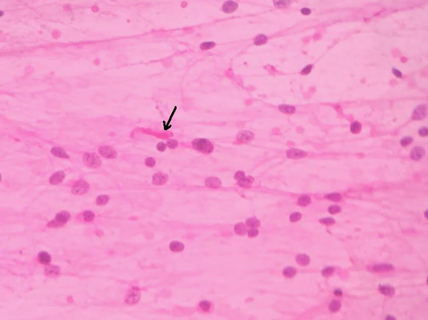
Eosinophilic Granular Bodies:
- Brightly eosinophilic granular clumps/ globoid deposits.

For those of you who are still thinking about "What a squash preparation is?"
- In squash preparation, a small piece of tissue is placed on one end of a slide. Another slide, right angles to the first slide is used to compress, squash and spread the tissue (length wise) over the first slide.
- It is then fixed, stained and examined under a microscope.
- Used in the intra-operative diagnosis of brain tumours.
Subscribe to:
Posts (Atom)