Thursday 27 December 2018

Ganglion cells - They will find you, before you find them😀

Ganglion cells are neuronal cell bodies.

They are identified by their ....

  • Large size
  • Polygonal shape
  • Large eccentrically placed vesicular nuclei
  • Prominent eosinophilic nucleolus
  • Abundant eosinophilic cytoplasm


Saturday 22 December 2018

Pilocytic astrocytoma - Squash preparation

Pilocytic Astrocytoma:

  • Most common astrocytoma in children.
  • Most common sites: Cerebellum, Floor of 3rd ventricle, thalamus, hypothalamus.


Most striking feature: Hair like (pilocytic processes) seen in the background.

Cells are monomorphic (uniform in size and shape) with ovoid bipolar bland appearing nuclei.
Many Rosenthal fibres and Eosinophilic Granular Bodies will be seen.

Rosenthal Fibres:
  • Brightly eosinophilic, thick, glassy cork-screw shaped/ carrot shaped srtuctures.


Eosinophilic Granular Bodies:
  • Brightly eosinophilic granular clumps/ globoid deposits.


For those of you who are still thinking about "What a squash preparation is?"
  • In squash preparation, a small piece of tissue is placed on one end of a slide. Another slide, right angles to the first slide is used to compress, squash and spread the tissue (length wise) over the first slide.
  • It is then fixed, stained and examined under a microscope.
  • Used in the intra-operative diagnosis of brain tumours.

Wednesday 19 December 2018

Diagnostic indicators of squamous cell carcinoma

Diagnostic indicators of squamous cell carcinoma:

1. Overlying epidermal dysplasia or tumour arising from the epithelium.

  • Squamous cell carcinoma, as the name indicates arises from squamous epithelial cells, the cells in the spinous layer.


Picture shows tumour arising from the overlying epithelium.

2. Single cell/ individual cell keratinisation. 

  • An important feature of squamous cells is to produce keratin, which is seen as an intense hyaline appearance.


Picture shows individual cell keratinisation - seen as dense eosinophilic area in the cytoplasm. Note the malignant nuclei.

3. Keratin pearl formation:

  • Tumour cells enclosing masses of anucleate keratin (Reminds me of a rose bud!!)
  • Seen in well differentiated tumours.


Picture shows keratin pearl - yellow circle;  resembles a rose bud!!
Individual cell keratinisation: green circle.

4. Intercellular bridges:


  • As these are epithelial cells and are connected to each other by cell-cell junctions (desmosomes), these can be seen in a tumour arising from squamous cells.


  • In a poorly differentiated tumour, where keratin pearls are absent and individual cell keratinisation may not be prominent, the only feature which helps in a morphological diagnosis is intercellular bridges.
  • These are better seen in areas of the tumour where there is oedema of the stroma.


In picture, very fine small lines are seen in between two tumour cells, connecting them.

Well differentiated squamous cell carcinoma is the most difficult diagnosis in surgical pathology!!

Tuesday 18 December 2018

Periodic Acid Schiff (PAS) staining

Tissue carbohydrates are of two types - Glycogen and Mucin (mucopolysaccharides).

Periodic Acid Schiff (PAS) stain:Indicates presence of glycogen in tissue

C-C bonds in carbohydrates (Eg:Glycogen) ----> Oxidised by periodic acid ----> Aldehyde ------> Schiff's reagent (Fuschin-sulphurous acid) ------> Alkyl sulphonate (magenta coloured compound).

Used in the diagnosis of:
1. Glycogen storage disorders


Liver biopsy (PAS stain) - demonstrating glycogen

2. Glycogen containing tumours - Clear cell renal cell carcinoma and Ewing sarcoma.

An important feature of glycogen: The ease of digestion with amylase; i.e Glycogen is digested by amylase; while mucin is not.
Thus, Glycogen is PASD sensitive while mucin is PASD resistant.
                    * Most commonly used amylase is ptyalin (salivary amylase).


Other structures highlighted by PAS stain (and of practical importance):
1. Basement membrane: 

  • Invasion of tumours especially squamous cell carcinoma of skin, cervix.
  • Thickening of basement membrane in diseases like DLE etc.
  • Renal biopsies: Capillary,  glomerular and tubular basement membranes

2. Fungal cell wall: 

  • PAS highlights the cell wall of living fungi (incontrast to Grocott's Methanamine Silver - GMS which highlights both living and dead fungi).

3. Renal biopsies - To differentiate the various glomerular diseases with organised deposits (in combination with Jones silver stain)

4. Neutral mucin

Sunday 16 December 2018

Acute inflammation

Acute inflammation consists of vascular reaction and cellular response.

Vascular reaction - three important steps - vasodilation, increased vascular permeability and emigration of leukocytes.

Emigration of leukocytes (neutrophils) involves margination,  rolling,  and  adhesion  to  endothelium. 

Here is a picture of MARGINATION OF NEUTROPHILS...
This is seen in an area adjacent to a focus of brain abscess..

Saturday 15 December 2018

Basics of H&E staining


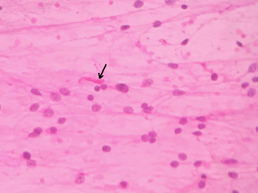
  • Hematoxylin and Eosin staining is the most common staining technique used in histopathology.
  • Displays the underlying tissue morphology with good contrast.
Basic concepts:
Why do dyes stain specific elements of cells and tissue?
  • Dyes demonstrate an affinity for molecules within cells and tissue.
  • Affinity is the result of attractive forces between dye molecules and molecules within the tissue.
Dye-Tissue interactions are facilitated by Vander Waal's forces, coulombic forces, hydrogen bonding and covalent bonding.

Haematoxylin: It is a basic dye and hence stains the acidic/ basophilic structures.
Nucleus/ nucleoplasm/ organelles that contain RNA - ribosome, ER etc


Eosin: It is an acidic dye and hence stains the basic/ acidophilic structures.
Cytoplasm/ cell wall/ extracellular fibres

In figure, blue staining --> hematoxylin, pink staining --> eosin

Quiz 2 - Cervical Cytology part 2

Loading…